

Ligaments of the Knee – Anatomy and Function
The knee joint is stabilized by several ligaments, which are strong bands of connective tissue that connect bones to other bones. These ligaments play a crucial role in providing stability and support to the knee during movement. Here's an overview of the major ligaments of the knee, along with their anatomy and functions:
1. Anterior Cruciate Ligament (ACL):
- Anatomy: The ACL is located in the centre of the knee joint and runs diagonally from the back of the femur to the front of the tibia.
- Function: The ACL prevents excessive forward movement of the tibia in relation to the femur and provides stability to the knee during activities such as pivoting, cutting, and jumping.
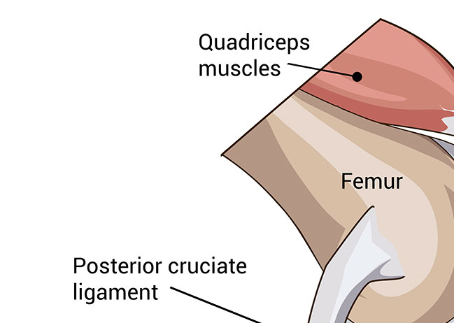
2. Posterior Cruciate Ligament (PCL):
Anatomy: The PCL is located in the back of the knee joint and runs diagonally from the front of the femur to the back of the tibia.
Function: The PCL prevents excessive backward movement of the tibia in relation to the femur and helps stabilize the knee during activities such as deceleration and landing from a jump.
3. Medial Collateral Ligament (MCL):
Anatomy: The MCL is located on the inner side of the knee joint and connects the medial (inner) aspect of the femur to the medial aspect of the tibia.
Function: The MCL provides stability to the inner side of the knee and helps prevent excessive inward (valgus) movement of the knee joint.
4. Lateral Collateral Ligament (LCL):
Anatomy: The LCL is located on the outer side of the knee joint and connects the lateral (outer) aspect of the femur to the head of the fibula.
Function: The LCL provides stability to the outer side of the knee and helps prevent excessive outward (varus) movement of the knee joint.
Lateral view of the knee
In summary, the ligaments of the knee work together to provide stability and support to the joint during various movements and activities, helping to prevent excessive motion and potential injuries.
